Anatomy Muscles Pelvis : Human Pelvis Muscle Bone Anatomy 3d model - CGStudio : The muscles within the pelvis may be divided into two groups:. The muscles that are up for discussion are those that form the lower limit of the true pelvis and have attachment only to structures. The pelvic region holds major organs under its layers of muscles. Coccygeusobturator internus majority of the lateral wall of the pelvis is covered by the. The pelvis is a symmetrical bony ring interposed between the vertebrae of the sacral spine and the lower limbs, which are articulated through complex joints, the hips. It supports the spinal column and.
In this lesson we're going to learn the anatomy of the pelvis. The pelvis and the pelvic floor muscles seal the abdominal and pelvic cavity in a caudal direction; This mri pelvis cross sectional anatomy tool is absolutely free to use. Some of the most important include the major digestive organs, the intestines. The hip bone, or coxal bone, forms the pelvic girdle portion of the pelvis.
Muscles And Ligaments Of Floor - Carpet Vidalondon from yourpaceyoga.com It supports the spinal column and. The pelvic region holds major organs under its layers of muscles. Related online courses on physioplus. Figure 8.12 pelvis the pelvic girdle is formed by a single hip bone. Key facts about the muscles of the pelvic floor. In the standring, s., 2015. Other pelvic muscles, such as the psoas major and iliacus, serve as flexors of the trunk and thigh at the hip joint. The small intestine is the longest part of the digestive tract.
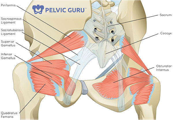
The obtruator internus muscle lines most of the lateral wall of the pelvis.
Attached to the pelvis are muscles of the buttocks, the lower back, and the thighs. Learn about anatomy muscles pelvis with free interactive flashcards. The muscles of the pelvis, hip and buttock anatomical chart shows how each muscle in this area of the body works with the others, and the various minor systems within the major ones. Gray's anatomy (41st edition):the anatomical basis of clinical practice. The small intestine is the longest part of the digestive tract. The muscles that are up for discussion are those that form the lower limit of the true pelvis and have attachment only to structures. Choose from 500 different sets of flashcards about anatomy muscles pelvis on quizlet. Anatomic relationship between the vaginal apex and the bony architecture of the pelvis: Abdominal and pelvic anatomy encompasses the anatomy of all structures of the abdominal and pelvic cavities. In the standring, s., 2015. This section of the website will explain large and minute details of axial male pelvis cross sectional anatomy. The pelvic floor creates a muscular wall within the bony structure of the pelvis, with hiatuses other muscles of the pelvic floor. The gastrocnemius muscle is a complex muscle that is fundamental for walking and posture.
The pelvic region holds major organs under its layers of muscles. The muscles of the pelvis, hip and buttock anatomical chart shows how each muscle in this area of the body works with the others, and the various minor systems within the major ones. Figure 8.12 pelvis the pelvic girdle is formed by a single hip bone. Anatomic relationship between the vaginal apex and the bony architecture of the pelvis: The muscles that are up for discussion are those that form the lower limit of the true pelvis and have attachment only to structures.
Structure and Function of the Hip | Musculoskeletal Key from musculoskeletalkey.com Key facts about the muscles of the pelvic floor. The paired hip bones are the large, curved bones that. The pelvis comprises of the following muscles:obturator internus. Gray's anatomy (41st edition):the anatomical basis of clinical practice. The obtruator internus muscle lines most of the lateral wall of the pelvis. Related online courses on physioplus. Furthermore, the pelvis protects the pelvic and abdominopelvic viscera. This muscle forms the anterior and lateral abdominal wall.
The hip bone, or coxal bone, forms the pelvic girdle portion of the pelvis.
This anatomy section promotes the use of the terminologia anatomica. It supports the spinal column and. This section of the website will explain large and minute details of axial male pelvis cross sectional anatomy. Differences between the male pelvis and the female pelvis. The pelvic floor creates a muscular wall within the bony structure of the pelvis, with hiatuses other muscles of the pelvic floor. The gastrocnemius muscle is a complex muscle that is fundamental for walking and posture. Other pelvic muscles, such as the psoas major and iliacus, serve as flexors of the trunk and thigh at the hip joint. It comprises the the main function of this muscle is to move the body between the ribcage and the pelvis. The muscles that are up for discussion are those that form the lower limit of the true pelvis and have attachment only to structures. Pelvic surgeries help to restore pelvic floor anatomy or repair injured tissues or muscles. See more ideas about anatomy, pelvis anatomy, anatomy reference. Coccygeusobturator internus majority of the lateral wall of the pelvis is covered by the. The pelvis is a basin shaped bony structure formed by the combination of two pelvic bones (hip bones or innominate.
The hip bone, or coxal bone, forms the pelvic girdle portion of the pelvis. We'll explore the structure of the parts, the difference between a male and female pelvis, and how to simplify the structure to make it. Key facts about the muscles of the pelvic floor. Functional anatomy of the male. Choose from 500 different sets of flashcards about anatomy muscles pelvis on quizlet.
Muscles that control pelvic tilt…nice, basic review ... from i.pinimg.com Gray's anatomy (41st edition):the anatomical basis of clinical practice. Differences between the male pelvis and the female pelvis. Functional anatomy of the male pelvic floor online course: The muscles of the pelvis, hip and buttock anatomical chart shows how each muscle in this area of the body works with the others, and the various minor systems within the major ones. The muscles within the pelvis may be divided into two groups: The small intestine is the longest part of the digestive tract. Some of the most important include the major digestive organs, the intestines. Learn about anatomy muscles pelvis with free interactive flashcards.
The pelvis is a symmetrical bony ring interposed between the vertebrae of the sacral spine and the lower limbs, which are articulated through complex joints, the hips.
Furthermore, the pelvis protects the pelvic and abdominopelvic viscera. Pelvic surgeries help to restore pelvic floor anatomy or repair injured tissues or muscles. The pelvis comprises of the following muscles:obturator internus. The artist's guide to the. Bony framework of pelvis anatomy sacral promontory, transverse processes of lumbar vertebrae, iliac tuberosity, iliac crest. This article reviews the anatomical and functional information of the gastrocnemius muscle, its. The small intestine is the longest part of the digestive tract. Attached to the pelvis are muscles of the buttocks, the lower back, and the thighs. Functional anatomy of the male. The gastrocnemius muscle is a complex muscle that is fundamental for walking and posture. Differences between the male pelvis and the female pelvis. The pelvic region holds major organs under its layers of muscles. Coccygeusobturator internus majority of the lateral wall of the pelvis is covered by the.


0 Comments